“Alzheimer's disease and other neurodegenerative diseases, such as Parkinson's and frontotemporal dementia, are characterized by abnormal protein aggregation in the brain. If you disrupt these aggregates but cannot clear the debris due to blockages in the drainage system (lymphatic system), then little progress has been made. You must clear the protein drainage channels to truly solve the problem.” — Xie Qingping
Basic Information on Alzheimer’s Disease
Alzheimer's disease (AD) is a progressively developing neurodegenerative disorder with an insidious onset. Clinically, it is characterized by symptoms such as memory impairment, aphasia, apraxia, agnosia, visuospatial dysfunction, executive function deficits, and changes in personality and behavior. The incidence of Alzheimer’s is rising annually, making it a significant health issue for the elderly in China. Epidemiological studies indicate that from 2005 to 2015, the prevalence of dementia in the elderly was about 5%, with AD accounting for 65% of these cases. Currently, the prevalence of Alzheimer’s among those aged 65 and older in China is approximately 3.21%. The exact causes of Alzheimer’s remain unclear.
It is widely believed that Alzheimer’s disease results from an imbalance in the production and clearance of amyloid proteins, with the meningeal lymphatic system playing a role in amyloid clearance. However, the unique location of lymphatic vessels hinders their drainage efficiency. Previously thought to be absent in the central nervous system, lymphatic vessels were discovered in the meninges during research on T-cell pathways to and from the brain. These structures exhibit all the molecular characteristics of lymphatic endothelial cells, capable of transporting fluids and immune cells from cerebrospinal fluid (CSF) and connecting to deep cervical lymph nodes. Surgical removal of deep cervical lymph nodes can disrupt normal T-cell flow in the meninges, leading to cognitive impairment.
Traditionally, CSF was thought to be produced by the choroid plexus in to Overview of Alzheimer’s Disease in China and Its Social Impact
In recent years, to meet the diverse needs of elderly care, the government has vigorously supported the rapid increase of nursing institutions, establishing a diversified and multi-layered institutional care system that adequately addresses the living and caregiving needs of older adults. However, the medical and health service system specifically catering to the healthcare needs of the elderly is not yet fully established. Nursing homes operate independently from medical institutions; they do not provide comprehensive medical services, while hospitals focus solely on disease treatment. As a result, elderly individuals who fall ill are forced to navigate between their homes, hospitals, and nursing facilities, leading to delays in treatment and additional burdens on family members. The separation of medical care and elderly care has caused many ill elderly patients to rely on hospitals as de facto nursing homes, exacerbating the strain on medical resources.
Currently, there are over 46 million Alzheimer’s disease patients worldwide, with one new case emerging every three seconds. China accounts for approximately one-quarter of the global total, with about 300,000 new cases diagnosed each year. Aging is one of the most challenging issues globally, affecting many developed countries. Despite being in a developmental phase, China is facing similar challenges. Population aging refers to the dynamic increase in the proportion of elderly individuals as the number of younger people declines.
According to a press conference held by the Health China Action Promotion Committee on July 29, 2019, research indicates that the prevalence of dementia among the elderly population in China is around 5.56%. Based on this, it is estimated that over 13 million elderly individuals in China suffer from dementia. Due to rapid aging, this number could exceed 40 million by 2050. The onset of dementia not only severely impacts the quality of life and lifespan of the patients themselves but also places a heavy financial burden on families and society, making it an increasingly serious public health and social issue.
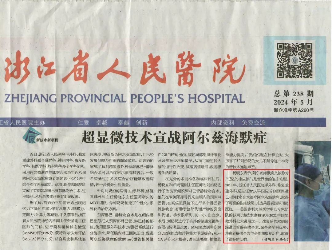
Hospitals like Hangzhou Qiushi Hospital and Hangzhou Huaguang Hospital have responded to the national call as early adopters of integrated medical and eldercare services in Zhejiang. Since 2014, they have focused on the treatment and research of geriatric diseases. Professor Xie Qingping, the medical leader at these hospitals and a visiting professor at the Cleveland Clinic in the U.S., has pioneered advanced supermicrosurgery techniques in the treatment of Alzheimer’s disease and other neurodegenerative disorders. His innovative deep cervical lymphovenous anastomosis technique, initiated in 2019, has treated nearly 300 patients with neurodegenerative diseases, including Alzheimer’s and Parkinson’s disease. Many patients have shown varying degrees of improvement in behavior, personality, and motor skills after surgery, with high family satisfaction reported.

What is Deep Cervical Lymphovenous Anastomosis (LVA) and Its Principles
Recent theories suggest that there are two lymphatic drainage systems in the brain (the glymphatic system and the meningeal lymphatic system), which serve as the only physical pathways for metabolizing harmful protein deposits. The lymphatic system not only has immune functions but also plays a role in transporting abnormal proteins from the brain. The principle of our surgical technique is to create a connection between corresponding lymphatic vessels and veins in the neck to reduce lymphatic pressure in deep brain tissue, accelerating the removal of Alzheimer’s-related metabolic waste (various proteins). This alleviates lymphatic edema in deep brain tissue, improves brain function, and promotes recovery of symptoms in Alzheimer’s patients.
To put it simply, the procedure bypasses blocked cervical lymph nodes, directly connecting the cervical lymphatic vessels to veins for quick drainage, thus efficiently removing beta-amyloid protein from the brain and enhancing cerebrospinal fluid flow to improve the patient’s condition and potentially restore brain nerve functions.
Scientific Principles and Explanations
Foreign scholars have researched the brain's lymphatic system. A study published by the University of Washington School of Medicine in Nature on April 28, 2019, indicated that the brain’s drainage system (i.e., meningeal lymphatics) plays a crucial but underappreciated role in neurodegenerative diseases, suggesting that repairing defective drainage systems could be key to unlocking potential Alzheimer’s treatments.
Dr. Jonathan Kipnis, a leading author and professor at the University of Washington, noted that "lymphatics are a sink." Amyloid plaques begin to form in the brains of Alzheimer’s patients long before symptoms like forgetfulness and confusion arise. For years, scientists have attempted to develop therapies to clear these plaques but with limited success. Aducanumab, a promising candidate drug, recently showed efficacy in slowing cognitive decline in one clinical trial but failed in another, puzzling scientists.
Kipnis discovered meningeal lymphatics as the brain’s drainage system in 2015. He later demonstrated in 2018 that damage to this system increases amyloid accumulation in mice. He speculated that the inconsistent performance of anti-amyloid drugs might be explained by differences in lymphatic function among Alzheimer’s patients. However, proving this hypothesis has been challenging due to the lack of tools to directly measure the health of human meningeal lymphatics.
Surgical Response from Professor Xie Qingping
When the production and clearance of Aβ protein are imbalanced, it leads to the accumulation of Aβ in the brain. There are primarily three ways to clear excess Aβ in the human body:
Aβ is degraded by various proteolytic systems. Extracellular enzymes from microglia and astrocytes, as well as intracellular systems such as lysosomes and the ubiquitin-proteasome pathway, can degrade soluble Aβ.
At the blood-brain barrier, Aβ can be cleared into the bloodstream through special transport systems in brain endothelial cells.
The third method, which is our focus, involves the clearance of Aβ through the lymphatic system and perivascular drainage pathways in the interstitial space. Since the discovery of the meningeal lymphatic system in 2015, researchers have noted its role in clearing Aβ.
Our surgical key is to reopen this clearance system, enabling it to function normally, which naturally leads to therapeutic effects for patients. This is why our surgical treatment of Alzheimer’s disease shows significant results.
The Cleveland Clinic, the second-largest medical center in the U.S., expressed "great shock" at Professor Xie's academic discoveries. In April 2023, he was appointed a visiting professor there and signed a long-term academic cooperation agreement. Currently, Hangzhou Qiushi Hospital collaborates with the Cleveland Clinic to align with international standards in lymphatic reconstruction treatments for Alzheimer’s disease. The first academic article has also been published in the U.S. journal PRS titled “Rewiring the Brain—The Next Frontier in Supermicrosurgery,” heralded by American peers as the next technical frontier in microsurgery.
Advantages of Surgical Treatment
Introduction to Lymphovenous Anastomosis (LVA) and Its Safety:
Lymphovenous anastomosis (LVA) is a minimally invasive surgical technique for treating lymphedema. It connects affected lymphatic vessels to adjacent veins, aiding lymphatic drainage and alleviating symptoms related to lymphedema. This technique is commonly used for lymphedema following breast cancer surgery and other causes.
The discovery and rapid development of LVA are credited to Japanese surgeon Mikinori Kono, who introduced it in the 1960s. By connecting blocked lymphatic vessels to nearby veins, this surgery enhances lymphatic drainage efficiency and alleviates patient symptoms.
As a mature surgical method, LVA is relatively safe, but requires a high level of anatomical knowledge and practical skills from the operating surgeon. According to professional organizations, there are currently fewer than 600 surgeons worldwide capable of performing supermicrosurgery, with very few experts. Professor Xie Qingping, as a pioneer of the latest cervical LVA theory, also conducts educational tasks in continuing education for lymphedema treatment, continuously providing updated operative techniques in China. In the near future, there is hope to establish a subspecialty in supermicrosurgery focused on intracranial lymphedema to serve more Alzheimer’s patients.
Effectiveness and Sustainability of Surgical Treatment
According to anatomical and natural science studies, the cervical lymphatic system serves as the only pathway linking the brain’s internal lymphatic system to systemic circulation. The process of treating blocked lymph nodes and draining them into veins rapidly releases the high pressure in the brain’s lymphatic system. This explains why patients often feel improved changes just one day or even a few hours after surgery.
Based on an article published by Professor Xie Qingping in the Chinese Journal of Microsurgery, the recovery of a patient post-surgery was as follows:
Patient Background:
An 84-year-old Alzheimer’s patient was diagnosed five years ago. Despite long-term medication, the condition worsened, leading to confusion, aggressive behavior, inability to eat, incontinence, immobility, severe forgetfulness, and visual-spatial impairment. An MRI prior to surgery showed age-related brain changes.
Surgical Decision and Approval:
Given the patient’s condition and the extent of daily life impairment, the doctor decided to attempt cervical deep lymphatic-venous anastomosis. After obtaining informed consent from the patient and family, the surgical plan was approved by the hospital’s ethics committee.
Surgical Process:
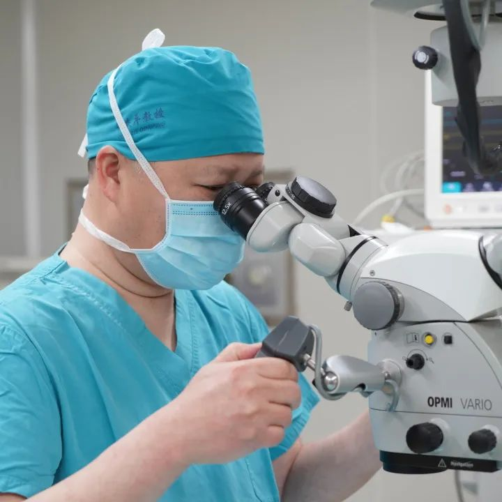
The surgery was performed using supermicrosurgical techniques under a surgical microscope magnifying 40-60 times, divided into two steps:
Lymphatic-Vein End-to-Side Anastomosis: After clamping to stop blood flow, the lymphatic vessel was trimmed and anastomosed end-to-side, ensuring no leakage and unobstructed blood flow.
Lymphatic-Venous End-to-End Anastomosis: The surgeon ligated the distal end of the vein and prepared the proximal end for opening, using 11-0 or 12-0 sutures to perform an equidistant anastomosis with the matched lymphatic vessel. After completing the anastomosis, the surgeon released the vascular clamp and checked for any leakage.
During the surgery, the surgeon noted that the fluorescent-stained lymph fluid flowed smoothly into the vein in a continuous manner, indicating the success of the operation.
Specific Steps:
Locating Lymphatic Vessels in the Neck: Preoperatively, Doppler ultrasound was used to detect the path of the accessory nerve lymph node chain in the cervical Va region and the condition of the lymph nodes in area V (the area between the posterior border of the sternocleidomastoid muscle and the anterior border of the trapezius muscle, with the lower limit defined by the lower belly of the thyrohyoid muscle and the upper limit as area V). The surgery was centered on the midpoint of the intersection of the posterior border of the sternocleidomastoid muscle and the external jugular vein, making an approximately 6 cm oblique incision along the posterior border of the sternocleidomastoid muscle.
Fluorescent Tracing and Anatomy of Lymphatic Vessels and Veins in Fat Tissue: During the surgery, 25 mg of indocyanine green was mixed with 0.8 ml of 0.9% saline (Dandong Yichuang Pharmaceutical) and diluted 5 times. 0.2 ml of the diluted solution was injected into the cervical venous opening via the carotid sheath. A laser lymph imaging device was used for lymphatic staining. Lymphatic vessels surrounding both sides of the accessory nerve lymph node chain were fluorescently marked, identifying lymphatic vessels with a diameter of approximately 0.2 mm, multiple for anatomical use. One lymphatic-venous end-side anastomosis and one end-to-end anastomosis were performed on the left side; one lymphatic-venous end-side anastomosis on the right side. The entire process was conducted under a 3D microscope, utilizing bipolar electrocoagulation for timely hemostasis.
Lymphatic-Venous Anastomosis: The lymphatic-venous anastomosis was performed under a binocular microscope at 40-60 times magnification.
Lymphatic-Venous End-Side Anastomosis: The diameter of the side-to-side anastomosis was 0.6 mm for the vein and 0.2 mm for the lymphatic vessel. An incision was made at a 30° angle to the vein's proximal end. The lymphatic vessel was trimmed again, resulting in lymphatic fluid leakage. The proximal end of the cut lymphatic vessel was left untreated, while the distal end was sutured using 11-0 or 12-0 sutures in an equidistant manner.
Lymphatic-Venous End-to-End Anastomosis: The diameter for the end-to-end anastomosis was 0.2 mm for both the lymphatic vessel and vein. The vein was managed by ligating the distal end, preparing the proximal opening, and performing an equidistant 4-suture end-to-end anastomosis with the matched diameter lymphatic vessel. After the anastomosis, the vein clamp was released, and bleeding was checked.
After the anastomosis, the incision and anastomosis site were checked for any leakage. Another 0.2 ml of diluted indocyanine green was injected at the cervical venous opening, revealing continuous lymphatic fluid flow into the vein on the laser lymph imaging device. After adequate hemostasis, a drainage tube was placed, and the incision was closed in two layers using 4-0 Vicryl sutures.
Changes in MMSE and MoCA Scores:
Preoperatively, the patient's MMSE score was 3, CDR score was 3, and MoCA score was 2, indicating severe dementia. However, at six months post-surgery, the patient's MMSE score rose to 20, CDR score decreased to 1, and MoCA score increased to 18, reflecting significant improvements in cognitive function and quality of life.
Successful cases of cervical deep lymphatic-venous anastomosis demonstrate its effectiveness in alleviating Alzheimer’s disease symptoms. This brings hope not only to Alzheimer’s patients but also opens new research avenues in the medical field, garnering widespread attention.
With ongoing technological optimization and expanded applications, cervical LVA is expected to be a significant breakthrough in Alzheimer’s treatment, bringing health and happiness to more patients and families.
On World Alzheimer’s Day, let us illuminate the light of memory together!


